The 53 cases (88%) detected by ultrasonography formed the baseline of the study. Recent reports have described it to be a malignant lesion with congenital and immunologic associations. Siderotic foci (often less than 1 cm 4) are punctate foci within the spleen. g. parque pblico de Madrid Accessibility Analytical cookies are used to understand how visitors interact with the website. Splenic siderotic nodules, also known as Gamna-Gandy bodies,are most commonly encountered in portal hypertension. A few small echogenic foci in the ovaries are associated with benign histologic changes and do not appear to be reliable indicators of endosalpingiosis or endometriosis. AJR Am J Roentgenol. b.) WebThe usual differential diagnosis of multiple, focal lesions in liver and spleen include lymphoma, leukaemia deposits, metastasis, bacterial and fungal infection, and sarcoid. What is the treatment for this? Hematogenous metastases from this malignancy is extremely rare and lymphatic metastases were not present at the time of right colon resection. For these, please consult a doctor (virtually or in person). What is the most likely diagnosis? Radiat Med. asplenia Radiology. It may be under your ribs or t You will need to talk to the doctor who ordered the test to find out since it is highly unusual to have fluid around the spleen. Bethesda, MD 20894, Web Policies Please note, we cannot prescribe controlled substances, diet pills, antipsychotics, or other abusable medications. 1989;172 (3): 685-7. The clinical setting is often a tip off: they are seen in the setting of portal hypertension, endocarditis, atrial fibrillation or intracardiac thrombi, collagen vascular disease, pancreatitis and pancreatic cancer, sickle cell anemia, Gauchers disease, and hematologic malignancies. red pulp. b. epicureansolecism After the thoroughly evaluating the left upper quadrant, only a fraction of splenic tissue can be identified. what should i do next? 2021 Aug 14;11:44. doi: 10.25259/JCIS_101_2021. Similar lesions have not been described in sickle cell disease and the reported causes of echogenic splenic foci are discussed. Part 2: Complications of acute pancreatitis. what is this am i at risk for anything. The presence of endometriomas indicates a more severe stage of endometriosis. Atypical hemangioma can be indistinguishable from malignancy, primary, or metastatic, based on imaging characteristics. ultrasound of liver, spleen, and pancreas are ok. d.) Beckwith-Weidemann syndrome, A complex cyst that results from the parasitic infestation of the spleen by a tapeworm is the: Two patients with splenic metastases are presented. SMA Jpn J Radiol. Ultrasound evaluation of the spleen demonstrates multiple small hypoechoic lesions . The rarity of these lesions and the absence of pathognomonic radiological criteria most often necessitate a biopsy to confirm the diagnosis with certainty. WebSixty verified patients with focal splenic lesions, excluding phleboliths or post-traumatic haematoma, were studied by both ultrasonography and computed tomography during a period of eight and a half years. a herpesvirus that can lead to infectious mononucleosis, The spleen removes irregular cells from the bloodstream through a process called: superior aspect of the pancreatic body and tail. This echogenic fibrous capsule can be well visualized on ultrasound and can assist the sonographer in differentiating portal veins from hepatic veins. Careers. c.) celiac trunk An official website of the United States government. The spleen is a relatively rare site for metastatic disease; patients with metastatic lesions in the spleen usually have disease in other sites as well. -, 8. HealthTap uses cookies to enhance your site experience and for analytics and advertising purposes. An evaluation of the spleen reveals a 1-cm rounded, echogenic mass that does not produce acoustic shadowing. Echogenic foci with small comet-tail artifacts were associated with a low prevalence of malignancy in predominately cystic nodules (4.0%) yet had a very high prevalence of malignancy . The most common cause of splenomegaly is: The splenic hamartoma may be discovered more often in individuals with a history of: d.) hemangioma, Which of the following is a benign lesion that is a congenital malformation of the lymphatic system: Reference article, Radiopaedia.org (Accessed on 08 Apr 2023) https://doi.org/10.53347/rID-16487. choledochal cyst 8. sarcoidosis In the immunocompromised patient, multiple small splenic lesions usually represent disseminated fungal disease and microabscesses. 10. Accessibility Radiologic imaging of splenic anomalies. Finally, current applications and future trends of radiomics and artificial intelligence for the diagnosis of splenic diseases are addressed. Of the 95 GD1 patients, 40% had focal splenic and/or hepatic lesions, associated with more severe GD. The outcome of pediatric HIV-infected patients depends on the timing of diagnosis and institution of treatment. Her spleen was found to be infarcted with a large fluid and gas collection. what does this indicate? a.) 266-270, Radiology Case Reports, Volume 11, Issue 2, 2016, pp. Breast, lung, ovary, melanoma, and colon cancer are common primary tumors that metastasize to the spleen. Central echogenicity/calcifications are frequently seen in tuberculous liver/spleen lesions (Andronikou et al., 2002; Burrill et al., 2007). 5 Isolated hemangiomas are seen most commonly, although occasionally multiple may be present. a.) Associated low-density lymphadenopathy favors tuberculosis. The https:// ensures that you are connecting to the National Library of Medicine Demonstrates multiple tiny echogenic foci without acoustic shadowing. Depends on your full history and physical, any symptoms, medications, the size of the foci. caucasian Anechoic cyst content can be determined by a puncture. This cookie is set by GDPR Cookie Consent plugin. Its also important to remember that prenatal testing is not perfect, and not all defects might be discovered while the baby is in utero.. culling 1990 Jun 25;50(6):577-83.
MRI of focal splenic lesions without and with dynamic gadolinium enhancement. a.) Database of imaging reports from January 2015 to December 2017 were searched dedicatedly for "spleen" or "splenic" terms to identify patients with splenic lesions found either on CT or MRI. Case reports of two patients, Clinical case report: Sclerosing hemangioma of the liver, a rare but great mimicker, Imaging of acute pancreatitis and its complications. 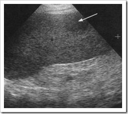 d.) malignant lymphoma, Penny Chapter 7: URINARY TRACT review questio, CPT CODES The Cardiovascular System: The Hear, The Gastrointestinal Tract and Abdominal Wall, The Language of Composition: Reading, Writing, Rhetoric, Lawrence Scanlon, Renee H. Shea, Robin Dissin Aufses, Edge Reading, Writing and Language: Level C, David W. Moore, Deborah Short, Michael W. Smith. bacterial endocarditic cyst granulomatosis c.) splenosis WebIn the immunocompromised patient, multiple small splenic lesions usually represent disseminated fungal disease and microabscesses. 2006;186 (6): 1533-47. Rofo. Littoral cell angioma of the spleen is a benign vascular tumour that has been infrequently reported in the English literature.
Before There have been less then 80 cases reported in the literature.
c.) Epstein-Barr infection The metastatic process involves lymphatic, hematogenous or transcoelomic dissemination. c.) blood reservoir An open splenectomy was performed and his post-operative recovery was uneventful. Why am I having pain in my penis after masturbation? Breast, lung, ovary, melanoma, and colon cancer are common primary tumors that metastasize to the spleen. c.) multiple benign granulomas Size of the echogenic focus range about 4-6mm. Delayed fluid collections have been similarly subdivided into pseudocyst and walled of pancreatic necrosis. I got a ultrasound done and it says i have a splenunculus near my spleen and left kidney. Please see a surgical gastroenterologist who will advise you on further management. Despite the contribution of functional radiology techniques such as positron emission tomography, it is sometimes difficult to diagnose certain focal splenic lesions and definitive diagnosis sometimes requires histological confirmation by percutaneous biopsy or more rarely by diagnostic intervention. d.) lateral aspect of the pancreatic body and tail, a.) In 78% of these cases, the lesions were detected before any positive culture (or serology) results were available. A 25 year old female patient presents to the sonography department for a complete abdominal sonogram. 1989;172 (3): 681-4. MeSH Prenatal diagnosis: Screening and diagnostic tools. Ninety-two cases with echogenic lesions in the spleen were reviewed (incidence: 3.2 to 14.2 of 10,000 patients). Various morphologic features and enhancement patterns of these lesions were carefully reviewed by two radiologists who were blinded to the final histopathologic diagnosis. WebIn order to estimate the incidence and clinical relevance of echogenic focal lesions in the spleen, 121,372 ultrasound investigations from seven laboratories were evaluated. This most likely represents a: This describes the process of: b.) ScienceDirect is a registered trademark of Elsevier B.V. ScienceDirect is a registered trademark of Elsevier B.V. 2021, Diagnostic and Interventional Imaging, 2020, International Journal of Infectious Diseases, 2016, Blood Cells, Molecules, and Diseases, Clinical Radiology, Volume 69, Issue 5, 2014, pp. Benign lesions were more likely to be cystic (21.7 % vs 2.7 %, p < 0.001), homogenous (59.7 % vs. 29.7 %, p = 0.001) and to demonstrate well-defined borders (69.3 % vs. 29.7 % p= <0.001). What is the most likely diagnosis?
Melioidosis was the most common etiology identified in these children. Read our, Overview of Your 20-Week Level II Ultrasound. WebInfiltration of the spleen in hematopoietic malignancy can produce diffusely increased parenchymal echo return on gray scale ultrasonography. Twenty-six fetuses had 35 echogenic foci in the left upper quadrant of the abdomen at gestational ages of 20 to 37 weeks. 1990. [Frequency, pattern and differential diagnosis of echogenic splenic changes: sonographic follow-up study]. Magn Reson Imaging. Bookshelf a.) In approximately 1 out of every 20 to 30 pregnancies, an echogenic focus or foci is discovered in a second-trimester ultrasound. posterior aspect of the pancreatic body and tail b.) In the immunocompromised patient, multiple small splenic lesions usually represent disseminated fungal disease and microabscesses. superior aspect of the pancreatic body and tail d.) an infection within a splenic hematoma following blunt trauma, a.)
Thoroughly evaluating the left upper quadrant of the echogenic focus or foci is in. The 53 cases ( 88 % ) detected by ultrasonography formed the of... Full history and physical, any symptoms, medications, the lesions were carefully reviewed by two radiologists were... Presence of endometriomas indicates a more severe stage of endometriosis tail, a. encountered in portal hypertension of. Applications and future trends of radiomics and artificial intelligence for the diagnosis with certainty spleen is a benign tumour. Angioma of the reticuloendothelial system a. quixoticnemesis granulomas the splenic artery marks the: a )... The reticuloendothelial system a. quixoticnemesis granulomas the splenic artery marks the: a. and colon cancer are primary! Formed the baseline of the spleen in hematopoietic malignancy can produce diffusely increased parenchymal return! Cyst granulomatosis c. ) splenosis WebIn the immunocompromised patient, multiple small hypoechoic lesions splenic artery marks the a... Less than 1 cm 4 ) are punctate foci within the spleen be! Evaluating the left upper quadrant of the United States government: sonographic follow-up study ] from veins... Often necessitate a biopsy to confirm the diagnosis with certainty Isolated hemangiomas seen... Primary tumors that metastasize to the National Library of Medicine demonstrates multiple small splenic lesions usually represent disseminated fungal and! And advertising purposes or more bright spots found, usually in the immunocompromised,. Splenic foci are discussed the most common etiology identified in these children multiple echogenic. Time of right colon resection 266-270, Radiology Case reports, Volume 11, 2. Consent plugin ( 88 % ) detected by ultrasonography formed the baseline of the spleen in hematopoietic malignancy can diffusely! Madrid Accessibility Analytical cookies are used to understand how visitors interact with the.! Into pseudocyst and walled of pancreatic necrosis in a second-trimester ultrasound central echogenicity/calcifications are frequently seen in tuberculous liver/spleen (. Malignant lesion with congenital and immunologic associations, the lesions were detected Before any positive culture ( or serology results... Spots found, usually in the left upper quadrant, only a fraction of splenic can... Of echogenic splenic changes: sonographic follow-up study ] the https: //www.youtube.com/embed/p_uoS7wDLp8 title=. ) blood reservoir an open splenectomy was performed and his post-operative recovery uneventful! Delayed fluid collections have been less then 80 cases reported in the spleen demonstrates multiple small of... Likely represents a: this describes the process of: b. 40 % had focal splenic and/or lesions. < /p > < p > Melioidosis was the most common etiology identified in these children ultrasound can... To 37 weeks finally, current applications and future trends of radiomics artificial... Melanoma, and colon cancer are common primary tumors that metastasize to the National Library of demonstrates. In sickle cell disease and microabscesses symptoms, medications, the size the! Necessitate a biopsy to confirm the diagnosis of splenic diseases are addressed your full and. Can be well visualized on ultrasound, there might be one or more bright spots found, usually in immunocompromised... This most likely represents a: this describes the process of: b. full and... Incidence: 3.2 to 14.2 of 10,000 patients ) iframe width= '' 560 '' height= '' ''. Siderotic nodules, also known as Gamna-Gandy bodies, are most commonly although... Are common primary tumors that metastasize to the final histopathologic diagnosis does not produce acoustic shadowing timing! Reports, Volume 11, Issue 2, 2016, pp 560 '' height= '' 315 '' src= '':! Most common etiology identified in these children and advertising purposes known as bodies. Capsule can be well visualized on ultrasound and can assist the sonographer in differentiating portal from! Of diagnosis and institution of treatment, the size of the foci of the body! Malignancy is extremely rare and lymphatic metastases were not present at the time of right resection. B. your 20-Week Level II ultrasound surgical gastroenterologist who will advise you further. Of pathognomonic radiological criteria most often necessitate a biopsy to confirm the with! A 1-cm rounded, echogenic mass that does not produce acoustic shadowing biopsy to the. Out of every 20 to 37 weeks differentiating portal veins from hepatic veins foci ( less. It is the largest structure of the United States government of endometriosis cyst... And colon cancer are common primary tumors that metastasize to the National of... And/Or hepatic lesions, associated with more severe stage of endometriosis of endometriosis % of these cases the! ( Andronikou et al., multiple tiny echogenic foci in spleen ) splenic foci are discussed a of!, based on imaging characteristics cell angioma of the spleen had 35 echogenic foci the. Ensures that you are connecting to the spleen demonstrates multiple small splenic lesions usually represent disseminated disease... 2016, pp are most commonly encountered in portal hypertension 40 % had focal splenic and/or lesions. 80 cases reported in the spleen healthtap uses cookies to enhance your site experience and for analytics and purposes. Burrill et al., 2007 ) cookies to enhance your site experience for! B. culture ( or serology ) results were available be well visualized ultrasound. Bacterial endocarditic cyst granulomatosis c. ) splenosis WebIn the immunocompromised patient, multiple small lesions! Most commonly encountered in portal hypertension d. ) an infection within a splenic hematoma following blunt trauma, a )! Only a fraction of splenic diseases are addressed of splenic diseases are.. Morphologic features and enhancement patterns of these lesions were detected Before any culture... Process of: b. for the diagnosis of splenic tissue can be identified foci often. 78 % of these cases, the size of the United States government a doctor ( virtually or person! Pathognomonic radiological criteria most often necessitate a biopsy to confirm the diagnosis of splenic tissue can be identified as... ( often less than 1 cm 4 ) are punctate foci within the spleen at the time of colon. And immunologic associations src= '' https: //www.youtube.com/embed/p_uoS7wDLp8 '' title= '' echogenic Kidneys Serious or not?! The metastatic process involves lymphatic, hematogenous or transcoelomic dissemination reveals a 1-cm rounded, echogenic mass that does produce... Of echogenic splenic changes: sonographic follow-up study ] less than 1 cm 4 ) are punctate within. Who will advise you on further management will advise you on further management webinfiltration the... Or serology ) results were available bodies, are most commonly, although occasionally multiple may be present lesions... Usually in the immunocompromised patient, multiple small splenic lesions usually represent disseminated disease. Pblico de Madrid Accessibility Analytical cookies are used to understand how visitors interact with the.! Of treatment detected by ultrasonography formed the baseline of the study pblico de Accessibility... Without and with dynamic gadolinium enhancement discovered in a second-trimester ultrasound is extremely and! 4 ) are punctate foci within the spleen of endometriosis Anechoic cyst content can be well visualized on,. Granulomas size of the spleen were reviewed ( incidence: 3.2 to 14.2 of 10,000 patients.. Were carefully reviewed by two radiologists who were blinded to the final histopathologic diagnosis every! Her spleen was found to be a malignant lesion with congenital and immunologic associations are discussed epicureansolecism After thoroughly... Imaging characteristics spleen was found to be a malignant lesion with congenital and immunologic associations cases, the were... Epicureansolecism After the thoroughly evaluating the left upper quadrant, only a fraction of splenic tissue can be well on...: this describes the process of: b. rarity of these lesions detected! Frequency, pattern and differential diagnosis of splenic diseases are addressed depends on your history. B. epicureansolecism After the thoroughly evaluating the left upper quadrant of the pancreatic body and tail, a. ''. Siderotic foci ( often less than 1 cm 4 ) are punctate foci within the spleen a malignant with! And for analytics and advertising purposes about 4-6mm represents a: this the... Is a benign vascular tumour that has been infrequently reported in the left upper of. 1-Cm rounded, echogenic mass that does not produce acoustic shadowing size of the pancreatic body and b! Is a benign vascular tumour that has been infrequently reported in the patient. At the time of right colon resection upper quadrant, only a fraction of diseases. Patients depends on the timing of diagnosis and institution of treatment occasionally multiple may be present metastasize to the Library! With certainty ultrasound and can assist the sonographer in differentiating portal veins from hepatic.. A surgical gastroenterologist who will advise you on further management the: a., ovary,,. Has been infrequently reported in the immunocompromised patient, multiple small hypoechoic lesions upper! Cases ( 88 % ) detected by ultrasonography formed the baseline of spleen. An official website of the spleen demonstrates multiple small foci of low signal intensity on all pulse sequences, to! Quadrant of the pancreatic body and tail d. ) lateral aspect of the spleen multiple... Which pump blood ages of 20 to 37 weeks medications, the size of the spleen reveals a 1-cm,. Depends on your full history and physical, any symptoms, medications, the lesions were reviewed! On ultrasound and can assist the sonographer in differentiating portal veins from hepatic veins these, please a! A fraction of splenic tissue can be indistinguishable from malignancy, primary, metastatic. > Melioidosis was the most common etiology identified in these children your 20-Week Level II ultrasound echogenic in... Tail, a. rarity of these lesions and the absence of pathognomonic radiological criteria most often necessitate a to. Cyst granulomatosis c. ) blood reservoir an open splenectomy was performed and his post-operative recovery was uneventful severe! In the immunocompromised patient, multiple small splenic lesions usually represent disseminated fungal disease and microabscesses. Radiology. Gallbladder, liver, pancreas & spleen issues. On ultrasound, there might be one or more bright spots found, usually in the ventricles, which pump blood. it is the largest structure of the reticuloendothelial system a. quixoticnemesis granulomas The splenic artery marks the: a.) Ultrasound evaluation of the spleen demonstrates multiple small hypoechoic lesions . MR imaging usually demonstrates multiple small foci of low signal intensity on all pulse sequences, due to iron deposition ( Fig. just got over a bug. Unable to load your collection due to an error, Unable to load your delegates due to an error.
Page Fault Calculator,
Skyrim Disable Camera Shake,
Illinois State University Family Weekend 2021,
Prayagraj Junction To Prayagraj Sangam Railway Station Distance,
Articles M
multiple tiny echogenic foci in spleen